

Adoptive Transfer EAE in SJL Mice
Recommended protocol for use with:
- Hooke Kit™ PLP139-151/CFA (native) Emulsion (cat. no. EK-0230), and,
- Hooke PLP139-151 in TC media (cat. no. DS-0161)
or,
- Hooke Kit™ [Ser140]-PLP139-151/CFA Emulsion (cat. no. EK-0120), and,
- Hooke [Ser140]-PLP139-151 in TC media (cat. no. DS-0121)
Summary
The adoptive transfer EAE model in SJL mice is recommended for study of compound effects on the effector phase of EAE, independent of the immunization process. Immunization occurs only in donor mice. Fully encephalitogenic cells are then transferred into recipient mice, where they induce EAE (effector phase of EAE).
The model is also used to test effects of compounds on differentiated Th1 or Th17 populations or cell trafficking. It can also be used to track effector cells in vivo.
In the adoptive transfer model, female SJL donor mice are immunized and used as a source of encephalitogenic T cells. Once donor mice have developed an immune response to [Ser140]-PLP139-151 (usually 10 days after immunization), they are sacrificed and their spleens and lymph nodes harvested.
Spleen and lymph node cells are cultured in the presence of [Ser140]-PLP139-151 to activate encephalitogenic T cells, which are then transferred to female SJL recipient mice to induce EAE.
Choice of PLP antigen
This protocol may be run with either native mouse PLP139-151 peptide, or serine-substituted [Ser140]-PLP139-151. For best results, the same peptide must be used for immunization of donor mice and for the later cell culture.
The native mouse PLP139-151 will produce more severe EAE; the [Ser140]-PLP139-151 peptide less severe. If unsure, our recommendation is to start with native mouse PLP139-151; this allows use of fewer donor mice (see protocol below).
In this protocol "PLP139-151", without further qualification, refers to either peptide, as chosen by the investigator.
Materials needed (per PLP139-151/CFA Emulsion kit, Hooke #EK-0120 or #EK-0230)
| Description |
|---|
| Hooke Kit™ [Ser140]-PLP139-151/CFA Emulsion (EK-0120) or Hooke Kit™ PLP139-151/CFA (native) Emulsion (EK-0230) |
| Hooke [Ser140]-PLP139-151 in TC media (DS-0121) or Hooke PLP139-151 (native) in TC media (DS-0161) |
| SJL donor mice, females, at 8 to 16 weeks old (Jackson Laboratory strain SJL/J) |
| SJL recipient mice, females, at 6 to 10 weeks old (The Jackson Laboratory strain SJL/J) |
| 70 µm cell strainers (BD/Falcon #352350) |
| TC flasks (Corning #430825) Note: We recommend Corning TC flasks. Quality of TC flasks is very important; cells do not develop equally well in flasks from all vendors. |
| Fetal bovine serum (FBS) for cell culture |
| RPMI 1640 |
| 1 M HEPES (Life Technologies, cat #15630080) |
| L–Glutamine–Penicillin–Streptomycin solution (Sigma #G6784) |
| MEM Non-Essential Amino Acids Solution (Life Technologies #11140-050) |
| MEM Sodium Pyruvate Solution (Life Technologies #11360-070) |
| 2-Mercaptoethanol (1000X, Life Technologies #21985-023) |
| Red Blood Cell Lysing Buffer (Sigma #R7757) |
| Trypan Blue solution (Sigma #T8154) |
| Sterile phosphate buffered saline (PBS) (standard formulation, pH 7.4, calcium-free, magnesium-free) |
| 70% isopropyl alcohol in spray bottle |
| 50 mL sterile polypropylene tubes |
| Pipettes - 5 mL, 10 mL, 25 mL |
| Media bottles |
| Petri dishes |
Protocol
The day of cell transfer is normally considered Day 0 of the study; therefore this protocol begins with immunization on Day -13.
Acclimate all mice to your facility for at least 14 days before starting.
If using native PLP139-151, use one donor mouse for each ~10 recipient mice.
If using [Ser140]-PLP139-151, use one donor mouse for each ~3 recipient mice.
Day -13: Immunize donor mice
On Day -13 immunize female SJL donor mice at four sites on the back with [Ser140]-PLP139-151/CFA emulsion (Hooke #EK-0120 or EK-0230). Inject 0.05 mL at each site (total of 0.2 mL of emulsion per mouse).
See Immunization of Mice for Generation of Encephalitogenic T Cells for detailed protocol.
Day -3: Tissue harvest, cell suspension preparation, culture set up
Do all cell prep aseptically in a biosafety cabinet. Keep cells cold (0 to 4 °C). Use cold media and keep cells on ice when practical.
Any FBS lot may be used for wash media, but the quality of FBS for the tissue culture media is critical, as all FBS lots are not equally able to support cytokine production in T cell cultures. Use a FBS lot selected to support maximum cytokine production by antigen-specific T cells.
Use a refrigerated centrifuge at 0 to 4 °C.
Prepare wash media (approximately 150 mL per 10 donor mice) by supplementing RPMI 1640 to reach the following concentrations:
- 2% FBS (lot of FBS is not critical in this preparation)
- 10 mM HEPES
Keep cold (0 to 4 °C).
Prepare tissue culture media (approximately 800 mL per 10 donor mice) by supplementing RPMI 1640 to reach the following concentrations:
- 10% FBS (selected for T cell culture)
- 2 mM L-glutamine, 100 IU/mL penicillin, 0.1 mg/mL streptomycin (1% of 100x solution)
- 1x MEM non-essential amino acids solution (1% of 100x solution)
- 1 mM sodium pyruvate (1% of 100 mM solution)
- 5.5x10⁻⁵ M 2-mercaptoethanol (0.1% of 55 mM solution)
- 10 mM HEPES (1% of 1 M solution)
Keep cold (0 to 4 °C).
Processing 3 to 5 donor mice at a time, euthanize mice, spray them with 70% isopropyl alcohol, remove inguinal and axial lymph nodes and spleens, and then place spleens and lymph nodes in a single Petri dish containing 10–15 mL of wash media.
Squash tissue from 4 to 5 mice in the Petri dish by pressing several times with the hard end of a 10 mL syringe plunger.
Place a fresh 70 µm cell strainer in a 50 mL tube. Collect all media and squashed tissue from the Petri dish into the cell strainer.
Using the soft end of a clean 10 mL syringe plunger, press the tissue through the strainer.
Carefully rinse the cell strainer into the tube with ~10 mL of wash media. (Lift one end of strainer off tube to let air escape; this helps avoid spilling material on the outside of the tube, losing cells.)
Repeat for all donor mice.
Keep cell suspension cold on ice until all donor tissues have been processed.
Spin down cells in 50 mL tubes for 10 minutes at approximately 300 g (do not put cells from more than 5 mice in one tube).
Carefully discard supernatant (avoid losing cells).
Resuspend the cell pellet in 2.5 to 3 mL per mouse of cold red blood cell lysing buffer, and keep cold for 4 to 5 minutes while red blood cells lyse.
While red blood cells are lysing, preload 35 mL of cold wash media into each of another set of 50 mL tubes, for use in the next step.
Keep cell suspension cold until cell lysis is complete. This is indicated by the solution becoming clear, bright red (before the lysis is complete, the cell suspension will be opaque red). This typically this takes 4 to 5 minutes. Watch carefully for color change in order to start the next step immediately (if unsure, stop lysis after 5 minutes).
Immediately after cell lysis is complete (within 10 seconds) add the previously prepared 35 mL of cold wash media to the cell suspension in each tube.
Spin down cells again for 10 minutes at approximately 300 g, then discard supernatant.
Resuspend the cells in ~5 mL wash media per mouse. Filter suspension through a fresh 70 µm cell strainer to remove clumps of dead cells.
Spin down cells again for 10 minutes at approximately 300 g, discard supernatant.
Resuspend the cells in ~5 mL tissue culture media per mouse. Again, filter suspension through a fresh 70 µm cell strainer to remove clumps of cells.
Count cells using Trypan Blue solution. Expected cell number is approximately 250 million cells from each donor mouse.
Transfer cells from all tubes into a single media bottle and dilute with tissue culture media to 3–3.5 million cells/mL.
Add PLP139–151 peptide (Hooke #DS-0121 or DS-0161) to the cell suspension to reach:
- If using native PLP139–151, 5 µg peptide/mL
- If using [Ser140]-PLP139-151, 20 µg peptide/mL
Note – IL-12 or IL-23 may be added to the cultures at 10 ng/mL. This will increase the number of Th1 and Th17 cells respectively and increase EAE severity.
Plate the cell suspension at 100 mL/TC flask (T150).
Culture for 70 to 72 hours, 37 °C, 5% CO2, humidified.
Day 0: Transfer cells to recipient mice
Collect cells from TC flasks into 50 mL tubes. Use 5 mL pipette to rinse flask bottoms as much as possible, but do not use scraper. There will be some leftover cells attached at the bottom; these are mostly macrophages and fibroblasts and do not need to be collected.
Spin down cells, 10 minutes at approximately 300 g.
Resuspend cells in approximately 2 to 3 mL PBS per flask.
Count cells using Trypan Blue solution. Expected cell number is approximately 90-120 million cells from each donor mouse.
Adjust cell concentration with PBS. For i.p. injection, adjust to:
- If using native PLP139–151, 20 to 30 million cells/mL
- If using [Ser140]-PLP139-151, 100 to 200 million cells/mL
-
Inject cell suspension into recipient mice i.p. at 0.2 to 0.3 mL/mouse targeting:
- If using native PLP139–151, 5 to 6 million cells/mouse
- If using [Ser140]-PLP139-151, 20 to 40 million cells/mouse
Note – If IL-12 or IL-23 was added to the cultures, inject 30-50% fewer cells.
Begin scoring mice 4 days after the cell transfer (see Appendix A).
Expected results
EAE onset is normally 6 to 9 days after the cell transfer in untreated mice.
The following illustrates typical results:
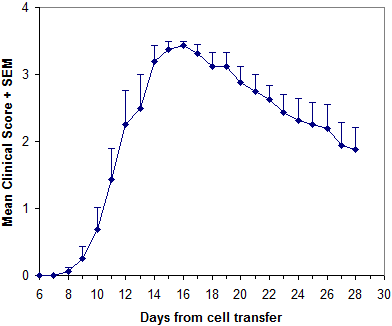
Hooke Kit™ [Ser140]-PLP139–151/CFA Emulsion (cat# EK-0120) was used for immunization of female SJL mice donor mice. Hooke [Ser140]-PLP139–151 in TC media (cat# DS-0121) at 20 µg/mL was used to stimulate encephalitogenic T cells for 3 days. Culture media was RPMI 1640 supplemented with FBS.
References
Thakker P et al, J Immunol 187:1986 (2011)
van der Veen RC et al, J Neuroimmunol 21:183 (1989)
McCarron R and McFarlin D, J Immunol 141:1143 (1988)
Appendix A - Mouse EAE Scoring Guide
Typically, EAE is scored on scale 0 to 5. Most researchers also give mice "in-between" scores (i.e. 0.5, 1.5, 2.5, 3.5) when the clinical picture lies between two defined scores.
The scoring method differs slightly depending on the stage of disease (onset/peak vs. recovery), for each individual mouse.
Reliable EAE scoring requires skill which comes after considerable experience. To avoid unconscious bias in scoring, we strongly recommend that mice should be scored blind, by a person unaware of which mice have received which treatment.
We recommend the following scoring guidelines for mice during onset and peak of EAE:
Mouse EAE scoring – onset and peak
| Score | Clinical observations |
|---|---|
| 0.0 | No obvious changes in motor function compared to non-immunized mice. When picked up by base of tail, the tail has tension and is erect. Hind legs are usually spread apart. When the mouse is walking, there is no gait or head tilting. |
| 0.5 | Tip of tail is limp. When picked up by base of tail, the tail has tension except for the tip. Muscle straining is felt in the tail, while the tail continues to move. |
| 1.0 | Limp tail. When picked up by base of tail, instead of being erect, the whole tail drapes over finger. Hind legs are usually spread apart. No signs of tail movement are observed. |
| 1.5 | Limp tail and hind leg inhibition. When picked up by base of tail, the whole tail drapes over finger. When the mouse is dropped on a wire rack, at least one hind leg falls through consistently. Walking is very slightly wobbly. |
| 2.0 | Limp tail and weakness of hind legs. When picked up by base of tail, the legs are not spread apart, but held closer together. When the mouse is observed walking, it has a clearly apparent wobbly walk. One foot may have toes dragging, but the other leg has no apparent inhibitions of movement. - OR - Mouse appears to be at score 0.0, but there are obvious signs of head tilting when the walk is observed. The balance is poor. |
| 2.5 | Limp tail and dragging of hind legs. Both hind legs have some movement, but both are dragging at the feet (mouse trips on hind feet). - OR - No movement in one leg/completely dragging one leg, but movement in the other leg. - OR - EAE severity appears mild when picked up (as score 0.0-1.5), but there is a strong head tilt that causes the mouse to occasionally fall over. |
| 3.0 | Limp tail and complete paralysis of hind legs (most common). - OR - Limp tail and almost complete paralysis of hind legs. One or both hind legs are able to paddle, but neither hind leg is able to move forward of the hind hip. - OR - Limp tail with paralysis of one front and one hind leg. - OR - ALL of:
|
| 3.5 | Limp tail and complete paralysis of hind legs. In addition to: Mouse is moving around the cage, but when placed on its side, is unable to right itself. Hind legs are together on one side of body. - OR - Mouse is moving around the cage, but the hind quarters are flat like a pancake, giving the appearance of a hump in the front quarters of the mouse. |
| 4.0 | Limp tail, complete hind leg and partial front leg paralysis. Mouse is minimally moving around the cage but appears alert and feeding. Often euthanasia is recommended after the mouse scores 4.0 for 2 days. However, with daily s.c. fluids most C57BL/6 mice may recover to 3.5 or 3.0, while SJL mice may fully recover even if they reach score 4.0 at the peak of disease. When the mouse is euthanized because of severe paralysis, a score of 5.0 is entered for that mouse for the rest of the experiment. |
| 4.5 | Complete hind and partial front leg paralysis, no movement around the cage. Mouse is not alert. Mouse has minimal movement in the front legs. The mouse barely responds to contact. Euthanasia is recommended. When the mouse is euthanized because of severe paralysis, a score of 5.0 is entered for that mouse for the rest of the experiment. |
| 5.0 | Mouse is spontaneously rolling in the cage (euthanasia is recommended). - OR - Mouse is found dead due to paralysis. - OR - Mouse is euthanized due to severe paralysis. |
In the recovery stage of EAE, most mice will have a tail that is no longer limp but is not normal either; it feels rigid and is "hooked". The hind legs may start moving (pedaling), but the mouse cannot walk. Either change makes scoring difficult.
We recommend the following modifications to the above scoring criteria for these mice:
Mouse EAE scoring – modified
| Score | Clinical observations |
|---|---|
| 0.0 | When held by the base of tail, tail is somewhat “hooked” and rigid, but tail makes complete rotations around the body axis (“helicopter”). Mouse is healthy. No signs of wobbling. |
| 0.5 | Mouse appears normal but tail is “hooked” and rigid. Tail does not make complete rotations around the body axis (“helicopter”). Mouse is healthy. No signs of wobbling. |
| 3.0 | Mouse is found on its side (as described for score 3.5 above), but there is excessive hind leg movement. Mouse cannot walk. - OR - Mouse has a wobbly walk (as described for score 2.5 above), and is unable to take more than two steps without falling on its side. The mouse is unable to right itself. - OR - Mouse has poor movement in the hind legs (as described for score 2.5 above), and has partial front leg paralysis evidenced by head held lower than normal and mouse's inability to right itself when placed on its side. |
| All other scores | Subtract 0.5 from the score of all mice with either a rigid, “hooked” tail or pedaling of hind legs. |
Version: 2023-04-23
_150px.jpg)