

Histology Services
Hooke's highly experienced histology team offers a comprehensive set of histology, IHC, and pathology services.
We accept fixed and frozen samples, embedded tissue blocks, unstained or stained slides, or digital images for analysis.
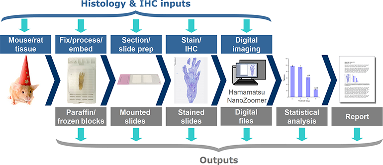
Available outputs include tissue blocks, mounted slides, and high-resolution digital images of your slides (Hamamatsu NanoZoomer). We can also perform statistical analysis and generate reports.
Histological staining
We routinely process and stain both formalin-fixed and cryopreserved tissue.
Hooke can decalcify fixed tissue, prepare optimal cutting temperature compound (OCT) blocks or formalin-fixed paraffin embedded (FFPE) blocks, and make slides.
Hooke's validated stains include:
- Alcian blue PAS
- Alizarin red S
- Congo red
- Hematoxylin and eosin (H&E)
- Jones' methenamine silver
- Luxol fast blue (LFB)
- Luxol fast blue (LFB) with PAS
- Miller's elastic stain
- Oil red O
- Periodic acid–Schiff (PAS)
- Phosphotungstic acid-hematoxylin (PTAH)
- Picrosirius red (PSR)
- Prussian blue
- Safranin O
- Silver stain
- Sirius red
- Sudan black B
- Tartrate-resistant acid phosphatase (TRAP)
- Toluidine blue
- Trichrome
- Verhoeff-Van Gieson (VVG)
- Von Kossa
Other stains can be obtained on request.
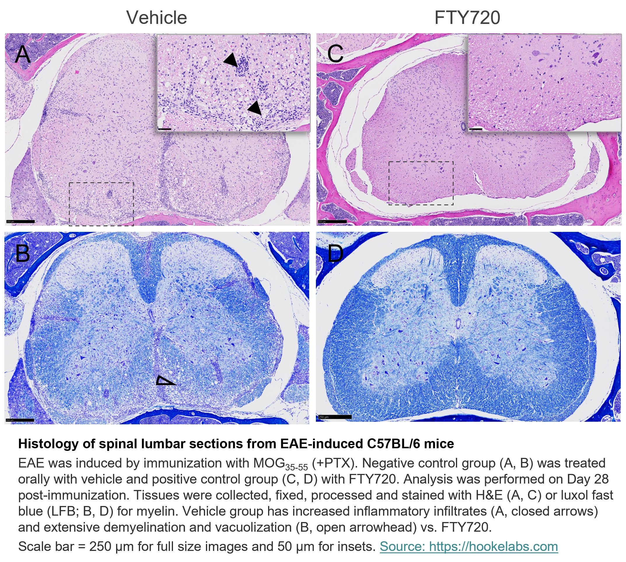
Histology of spinal lumbar sections from EAE-induced C57BL/6 mice
EAE was induced by immunization with MOG35-55 (+PTX). Negative control group (A, B) was treated orally with vehicle and positive control group (C, D) with FTY720. Analysis was performed on Day 28 post-immunization. Tissues were collected, fixed, processed and stained with H&E (A, C) or luxol fast blue (LFB; B, D) for myelin. Vehicle group has increased inflammatory infiltrates (A, closed arrows) and extensive demyelination and vacuolization (B, open arrowhead) vs. FTY720.
Scale bar = 250 µm for full size images and 50 µm for insets.
Multiplex immunohistochemistry (IHC) and immunofluorescence (IF)
Hooke has validated the following IHC marker antibodies:
- Adipophilin
- APP-beta amyloid
- CD11b
- CD3
- CD4
- CD45 (all isoforms, both CD45.1 and CD45.2 alloantigens)
- CD45R (B220)
- CD68 (F4/80)
- Collagen type I
- FOXP3
- Glial fibrillary acidic protein (GFAP)
- Iba-1
- Ki-67
- Myelin basic protein (MBP)
- Olig-2
- phospho-STAT1
- phospho-STAT3
- Smooth muscle actin (SMA)
We can also work with you to validate additional markers.
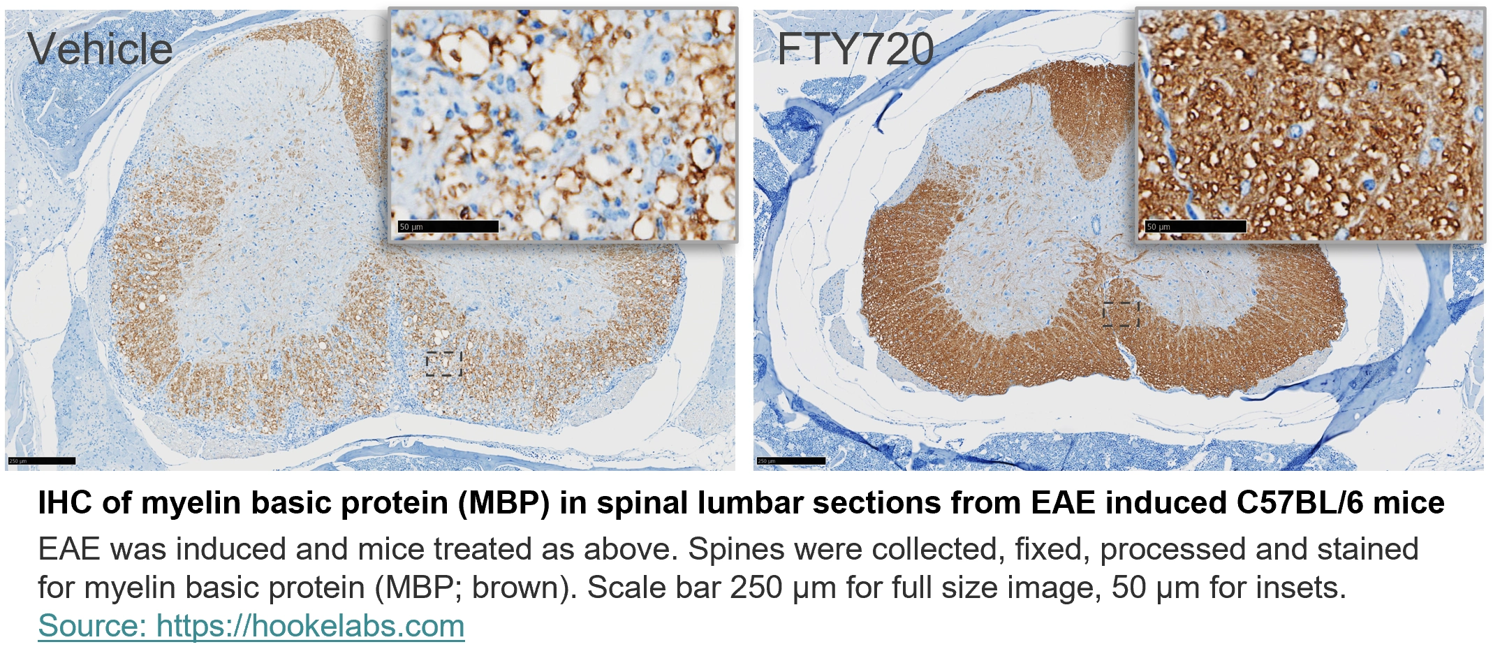
IHC of myelin basic protein (MBP) in spinal lumbar sections from EAE induced C57BL/6 mice
EAE was induced in C57BL/6 mice by immunization with MOG35-55 + PTX; analysis was on Day 28 after immunization. Treatments FTY720 (positive control, left) and vehicle (negative control, right) were administered prophylactically (from EAE induction), p.o. QD. Tissues were collected, fixed, processed and stained for myelin basic protein (MBP; brown). Scale bars 250 µm.

IHC of anti-phospho-STAT from CD4+CD45RBhigh-induced colitis in SCID model
Healthy (CD4 transferred mice; left) and colitis-induced (CD45RBhigh transferred mice; right) mice were sacrificed at 42 days post-colitis induction. Colons were cleaned, fixed, and immunohistochemically stained for anti-phospho-STAT3 (aP-STAT3, brown). Scale bar 2.5 mm for full size image, 50 µm for insets.
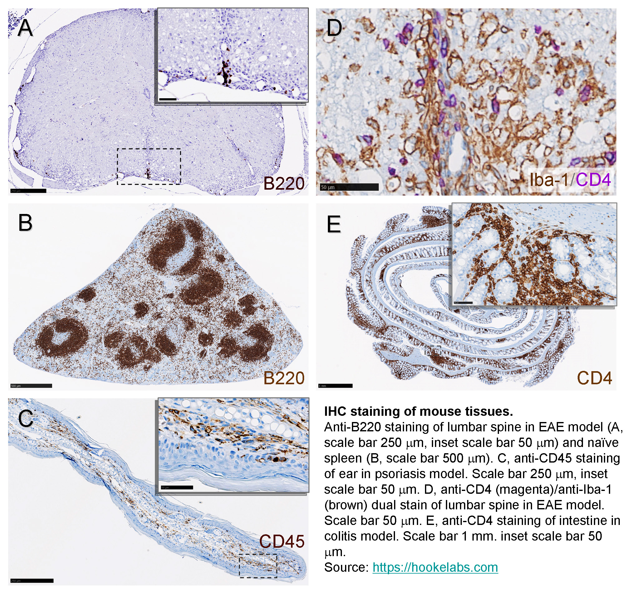
Pathological analysis
We offer pathological scoring of rodent spine, colon, kidney, spleen (including follicle counts), paw, and ear tissue (including dermal thickness measurement) for inflammatory and autoimmune disease models, including:
- Collagen-induced arthritis (CIA)
- Experimental autoimmune encephalomyelitis (EAE)
- Inflammatory bowel disease (IBD; colitis, Crohn's disease)
- Psoriasis
- Systemic lupus erythematosus (SLE)
Ordering and quotations
For a quotation or to order services, please contact us at .
_150px.jpg)